Video Library
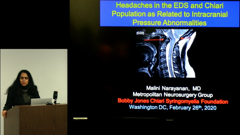
Headaches in the EDS and Chiari Population & Intracranial Pressure Abnormalities
DOCTORS COMMUNITY HOSPITAL - LANHAM, MD - FEBRUARY 26, 2020
The correct diagnosis and treatment of headache is a very complicated process that involves a lot of back-and-forth discussion between patient and provider. There are many different conditions that can cause headache, many of which neurosurgeons have to consider when diagnosing their patients.
Dr. Malini Narayanan of the
Metropolitan Neurosurgery Group gives an excellent and thorough overview of headache-causing disorders, with a special focus on pseudotumor cerebri, also sometimes called "idiopathic intracranial hypertension" (IIH) or "benign intracranial hypertension". Pseudotumor cerebri is a particularly challenging condition and especially troubling for surgeons because surgery is seen as a truly last resort.
Dr. Narayanan concludes her presentation by reminding us that a good doctor treats a disease, but a great doctor treats the patient. This is a particularly poignant point when discussing such a confusing medical landscape as
Chiari,
Ehlers-Danlos syndrome and related disorders.
This presentation was given at the February 26, 2020 Bobby Jones CSF patient and provider meeting in Lanham, Maryland. (2020)
Full Video Transcript:
[Dorothy Poppe]: So, I just want to welcome everyone here tonight. I really want to thank Dr. Henderson always for being such a supporter and he has so many things going on all the time, but he always makes time for the group and for his patients and we're very grateful to have him as one of our leaders.
Tonight, we're really excited to have his partner-- I'm going to let Dr. Henderson do the honors--so I'm going to hold off.
But-- I just want to say... I'd like everybody just to have a thought... We have a very dear friend, Shirley Cleaver, who is-- um-- has been diagnosed with Stage IV pancreatic cancer and if everyone would just take a moment to say a little prayer...and send some good thoughts her way I would really appreciate that, so—
[thoughtful silence]
And I just want her to know. So, um-- with that, I'm going to bring up Dr. Henderson.
[Dr. Henderson]: Thanks, Dorothy.
I'm very honored tonight to have Dr. Koby, my colleague, Dr. Rosenbaum, my new colleague, Dr. Narayanan, who I'll be introducing. And J.R. Bucklin, one of the foremost doctors of physiotherapy in the country. Dr. Pocinki, Dr. Affelbaum...and uh-- did I miss anyone? Any other doctors? And some of our extraordinary patient advocates. Um—so tonight, I'd like to introduce Dr. Malini Narayanan who's our new colleague extraordinaire.
Malini did a baccalaureate at the University of Massachusetts Amherst and then an engineering degree in electrical and computer engineering at M.I.T. Which-- I don't know what the "M" stands for-- I don't know if it's "Maryland" or--[laughter] M.I.T.-- whatever that is? And then a medical degree at Chicago. Her internship and residency at Harvard. And then fellowship in pediatric neurosurgery back at Chicago. We felt very lucky to get her to join our group. She's done a lot of research in mitochondria and some Chiari and spine work. And uh-- she's very well rounded. She's won prizes as most compassionate doctor of the year, top doctor, top neurosurgeon, top--one of the leading physicians in the world, and one of the Washingtonian's top doctors.
So, welcome Malini. Malini is going to talk about two topics that are really confusing and they've-- recently over the last couple of years, I think there's been a lot of confusion about the issues of intracranial hypertension, and spinal headaches which can occur separately, or even together. And they can also be misdiagnosed because there are a lot of other conditions that can mimic intracranial hypertension. So, Malini-- Dr. Narayanan is going to try to clarify these issues for us. So, welcome, Dr. Narayanan.
[Dr. Narayanan]: (Thank you so much)
[clapping]
[From audience] Can I just ask that you project?)
[Dr. Narayanan]: Stand straight. That's what I remember Dr. Henderson saying.
[Dorothy Poppe]: No, we have a web conference going tonight with lots of people.
[Dr. Narayanan]: Wonderful.
[Dr. Narayanan]: Here, let me just bring this back to the front.
[Dr. Narayanan]: I think because it's...it's touchscreen, so you can just go forward.
But Dr. Henderson, thank you so much for such a generous introduction. And I likewise feel honored to be joining Metropolitan Neurosurgery Group with you and Dr. Rosenbaum. It's a real delight. I'm also honored to be giving this talk today.
I mean, this is a very complex topic of headaches. And, you know, there's many appropriate titles-- I think I changed it a couple of times and I settled in on "Headaches in the EDS and Chiari Population as Related to Intracranial Pressure Abnormalities". And I think one of the most fun aspects of this talk is really going to be the discussion and hearing what people have to say about it.
I wanted to start from the top you know, just kind of stepping back because sometimes when we get so super-specialized, we think of only one type of headache over, over and over. So just a very quick slide on the broad topics we, as neurosurgeons, think about-- Vascular etiology: AVM, aneurysm, even trigeminal neuralgia, where there's an artery pressing on cranial nerve 5, causing excruciating facial pain and leading to headaches, as well. Traditionally, in the vascular set the onset when an aneurysm bleeds leads to a very sudden onset--severe headache. Excruciating. The worst kind of headache, ever.
In an AVM, it can be just the mass effect if-- and this is an "arteriovenous malformation"-- if it doesn't rupture or cause a bleed. Trauma. I think-- I've seen many EDS patients particularly where all of their symptom’s kind of start after a particular trauma. Whether it be a skiing accident, or athletics, um-- and so that's another aspect of headache.
Intracranial bleed, venous occlusion-- which we're going to get into with idiopathic intracranial hypertension Tumors: meningioma is just one that came to my mind, but of course, there's a whole slew of tumors that cause mass effect within the head that then causes many headaches. And that's where the name "psuedotumor cerebri"--that is, "false tumor"-- it acts like a mass, because you have this horrible headache. Congenital: Chiari 1 malformation, hydrocephalus. Infection: meningitis, sinusitis. Thinking about the headache in the front. And meningitis, you're thinking about "nuchal rigidity" or a very stiff neck. Systemic: hypertension, facial muscle contraction, TMJ, toothache, opiate withdrawal, even...CSF-related: occult CSF leak, idiopathic intracranial hypertension, hydrocephalus.
So, the white is kind of highlighted into what we look at, or what we think about a lot. Epidemiology of idiopathic intracranial hypertension-- also known as pseudotumor cerebri, also known as benign intracranial hypertension. The incidence in the general population is 1-2 over 100,000. Incidence in obese women of child-bearing age is 19-21 out of 100,000-- almost making it 10 times more. Peak incidence is in the third decade [of life], 40% occur in children, with 90% occurring in ages 5-15. Children under 12: the male to female incidence is almost equal. But post-pubertal, you almost have 90% women who get pseudotumor cerebri or IIH. Six to 24% --and this is quite a range—of patients with IIH have Chiari 1 malformation...and actually, vice versa. A diagnosis of Chiari 1 malformation can bring on in about 6% to 10% of IIH. So, it can be tricky.
I just wanted to get another big-picture slide, thinking about ICP versus volume and this is called the Monro-Kellie doctrine. The idea is that we, and this was a static model, really when it was first developed-- where they studied cadavers or in animals, this is a fixed volume of our skull and as we inject more volume into that skull, there is a very acute increase in the pressure, beyond a certain point in volume.
So, you see that right about here. And, so that's the challenge even with pseudotumor cerebri-- there's diurnal variation, throughout the day-- so sometimes, even if you're fairly convinced clinically that a patient has this problem and you do a single point lumbar puncture, and you feel like the pressure is not as high as it should be, that may be warranted: to do the lumbar puncture again at another time.
The dynamic aspect of ICP is really enormous. I mean, you're talking about ionic channels that work at cerebrospinal fluid production. You're looking at absorption at a molecular level. You're looking at flow--venous congestion, which is the blood flow draining out of the brain, the blood flow draining in. All of these work together in a complex, dynamic way to come out with an intracranial pressure that could either be pathologic--that is high-- above 20-25--or normal. And it's working constantly. When we cough-- all of us-- when we cough or when we bear down, our ICP's transiently go extremely high, but nothing happens to us.
And so, as I alluded, raised intracranial pressure is the diagnosis of idiopathic intracranial hypertension. It's usually >20, but a pressure greater, even far beyond, like 25-- has to have no evidence of mass lesion on your imaging. No evidence of lesions such as a large stroke, or any mass-occupying structure or abnormality. No hydrocephalus, so that means the ventricles are not large. No infection in the cerebrospinal fluid. When you do that lumbar puncture, you want to send off that CSF for an extensive profile. And no other contributing factors.
Pretty basic-- I think most people have had this, but your lumbar puncture: a needle into your back, between the spinous processes. That would be right along here... When you put that in, you don't want to lose a lot of CSF, you want to make sure, for example, if that patient in this case is awake, if they're breathing fast and exhaling, then it's going to artificially bring your pressures low.
Similarly, if you do it under intubation, or under conscious sedation, you want to make sure that your CO2 is not very low. It's in a normal state, between 35-45.
So, there's a lot of factors that go in--In this case, this patient is sitting up, so your pressure would actually be inaccurate, and you want the patient lying recumbent when you measure ICP.
The bolt is another way because with a lumbar puncture, you only get a single value of pressure, and with a bolt, you can get a few days. Usually the pressure monitor is placed right above the brain, underneath the dura. And you can get for many days, the pressure, but again, it can also have variations based on bearing down, constipation, any of those issues can make the ICPs either go very high and rarely can go very low unless you have an occult CSF leak.
So, as I said before, usually in the third decade, women greater than men in post-pubertal, headache in 94%. On exam or on symptoms, you're going to have visual blurring, eye pain or even loss of vision. You can have tinnitus, it can be pulsatile or whooshing noise. Normal radiologic studies, in terms of no mass-occupying lesion.
Obviously, if you have really high pressure sometimes you can have other findings like increased CSF spaces, especially where the pituitary gland resides. And it's called an "empty sella". You can have CSF pressure by lumbar puncture greater than 20. Normal CSF composition. And, essentially, a normal neurologic exam, except for, possibly, vision.
Interestingly, the headache, besides just being bilateral and behind the eyes, it can be pulsatile. It can also present as a migraine. There was a study of patients who were not improving on any medication regimen with migraines and these headaches presented as unilateral, and in these patients, more than 50% had elevated ICP when measuring the lumbar puncture in the opening pressure.
So, their take-home message was to consider, in a patient with a migraine, where it's not responding to any treatment, to consider lumbar puncture and rule out idiopathic intracranial hypertension.
You can have double-vision because of the cranial nerve 6 palsy. Transient visual loss, black outs, grey outs, blur outs, permanent visual loss. Nausea without vomiting in most cases. And there has been a number of studies, one recently from JHU about cognitive changes where there's language, memory and learning and executive function-- mostly in the memory and learning, 90% of the patients had some deficit in memory and learning.
So, the exam is actually getting a little more complex than to just say "absolutely normal". Furthermore, some of the symptoms can be the neck stiffness, torticollis-- more often in children. Low back pain, tinnitus-- a rushing noise-- and ataxia. Truncal ataxia mostly. Paresthesias in the hands and fingers. Dizziness in 30%. Fatigure, arthralgias, and reduced sense of smell. What is my neurosurgeon doing? Off-hand, I'd say you're suffering from an arrow through your head, but just to play it safe, I'm ordering a bunch of tests.
[laughter]
That's how it must feel for all of you when you come into our clinic. But now you can see that it can get-- even something like benign intracranial hypertension can get complex. Talked a little bit about that... So, factors linked to idiopathic intracranial hypertension... Trying to get the slide before...
Okay. There are many factors that influence idiopathic intracranial hypertension.
One paper kind of presented how to kind of decipher how strong this factor is. So, patients meet criteria for IIH, condition should be proven to increase ICP, treatment of the condition should be proven to improve IIH, and properly controlled studies should prove link.
And in looking at it from that point of view, obesity really qualifies as 4 out of 4 as a factor that really influences idiopathic intracranial hypertension. Cerebrovenous outflow obstruction which again, talking earlier about what influences intracranial pressure from an anatomic point of view, there is a backflow of blood being drained out of the brain, which then increases the pressure.
Strong link is hypervitaminosis A. And moderate link is oral contraceptives, Addison's disease, iron deficiency/anemia, polycythemia vera, uremia, lithium danazol and tetracycline. Possible link is Cushing's syndrome, minor head trauma and menstrual irregularities. And unlikely link is hyperthyroidism and steroid use. And no link is pregnancy.
I'm uh-- having fun with this touch screen as you can see...
So, really once you analyze for this and you feel like it's idiopathic intracranial hypertension, how many in the room have that condition? So, you probably remember the drill in terms of, you know, if your BMI is greater than 30, even 10% of weight loss can really reduce or reverse symptoms. And obviously, we would send for a nutritional consult with a specialist. That's if we have the time to do so and the visual deficits aren't progressive.
Medications: avoid at all costs opiate medication. Acetazolamide, metazolamide, Topamax, Lasix, high steroid use, and if there's a venous occlusion, anticoagulation.
"I have the solution to all of your health problems; all you have to do is change everything about the way you live."
[laughter]
How hard can that be? Stop the offending conditions. If you have the Vitamin A increased levels, uremia, tetracyclines. Stop it.
Minor procedure... at that point is serial lumbar punctures. Of course, it rarely happens, but it is a very scary list, you can have: cerebellar herniation, occult CSF leak, even an epidural hematoma on the spine, sciatica, weakness in legs, blood clot in the brain, and, rarely, uncal herniation.
And of course, all of these risks are rare. I've never seen it happen, but there have definitely been reported incidents and it's something that we think about when we're doing this.
We look at the head CT prior, and always think about, you know, make sure that there's no anticoagulation on board. And if there's an occult CSF leak, you can develop low-pressure headaches, which we'll talk about a little bit later.
If you've failed all the nonoperative therapy, and you have debilitating headaches, or progressive visual symptoms, then we really have to think about surgical options. At that point, the two options are a lumbo-peritoneal, or a ventriculo-peritoneal shunt. And I wanted to generalize it for people who may not be as exposed to shunts...
A shunt has CSF flow through a tube, go into a valve, and then drain into another part of the body. So, that's kind of a very broad way to think about them. In the lumbo-peritoneal, the CSF is siphoned off from the back and goes through a valve and then goes into the peritoneum, which is the belly/abdomen area.
The ventriculo-peritoneal shunt is from the brain, where a tube is placed in the ventricle. Very challenging because in patients with idiopathic intracranial hypertension, the area where you want to cannulate to drain off CSF is very, very small.
In fact, it may not be therapeutic enough, because that area is so small it's almost like trying to take fluid through a straw and over-suck and it just collapses. It's just not enough fluid to be able to bring the pressure down. You also have to oftentimes use stereotaxy in order to be able to cannulate the ventricle.
Consent process. Pretty scary, but certainly, we don't do that—and that's not the way it should be.
But it is hard. It's very complex, right? When you think about consenting for these procedures like a lumbo-peritoneal shunt or a ventriculo-peritoneal shunt and you're having debilitating symptoms. I want to talk a little bit about the shunt. The valve type is flow-dependent, pressure-dependent, position-dependent, anti-siphon, and usually there's a tapping reservoir. So, all of this is trying to-- you know, these are all different types of valves. You don't put them all in, you have to choose one.
The extracranial place, usually it's in the abdomen in the intra- peritoneal area, but it can be the lungs, the heart, the ureter, and the gall bladder. Very rare.
This is more a case in—certainly in pediatric cases-- where they have high pressure hydrocephalus, where it's imperative that they have a shunt. And in over 10-15 revisions, you end up running out distal places for the CSF to actually get absorbed.
Very interesting story... one of the 2nd round shunts... How many know the story about the engineer and the trio? So, one of the 2nd round valves and shunts that were developed I believe in the 1960s was a father whose son had trauma and was shunt-dependent and at that time, the shunt was giving enormous amounts of problems worked and put together an engineer with the neurosurgeon to develop a better shunt system and by the time they developed that, his own son didn't need it anymore.
And it kind of reminded me of all of you, who work so hard as patients to advance the field. And this is an example of that.
So, what are some of the risks? Because I just showed that scary comic picture of the consent. This is just a very fly-by, limited list and so when we think about surgery, you really are in a serious situation where our choices are less and less. And that's why we want to so much exhaust all the nonoperative therapies. Going up on the medications, trying steroids. If we feel obesity is a contribution, trying to get the weight loss. Trying to really core down on what the symptoms are.
Can you live with the headache? Are we doing okay? Is the vision not getting any worse? You know, how can we absolutely try to avoid surgery? Now, these surgeries are relatively safe, believe it or not. And have very low risk.
However, once it happens, it's like driving a car...There's no guarantee that every time you drive the car, there will never be an accident.
And in this case, when a risk manifests itself, everything right can be done, but there's just an inherent risk to any procedure. And in this case, infection is one of the risks, mostly in the CSF, very rarely in the brain if it's a ventriculo-peritoneal shunt.
Or in the extracranial site, such as the peritoneum. Abscesses, malfunction, brain and spinal bleeds. I kind of lumped the risks for both the shunts to make it easier. And even that can rarely cause paralysis or coma.
Overshunting with low pressure headaches, which can actually lead to bleeds over the brain. Shunt discomfort and overshunting can lead to an acquired Chiari malformation as well. Amazingly, even with a working shunt, sometimes your visual disturbances can persist and in that case--and part of that is because the reduction of the pressure has not transmitted all the way to the optic nerve. And so, in those cases, we may have to refer the patient to an additional procedure of an optic nerve fenestration.
So, now we're going to get into some cases. One case is sinus thrombosis. You can have a progressive global headache, personality change, loss of memory, had increase in leg weakness, urinary incontinence. Already, we know that this is not going to be pseudotumor cerebri or idiopathic intracranial hypertension but in some cases, there can be venous ouflow slowing, or a smaller venous obstruction that can cause some of those symptoms.
And in this case, we see a thrombosis of the straight and transverse sinus. Treated with enoxoparin, and recovered all the symptoms and recovered with an excellent neurologic exam. Interesting, sinus thrombosis is such a serious condition that it's one of the rare situations that even if you're initial CT with a sinus thrombosis shows some bleed, like brain bleed, you would still do full anticoagulation.
Here's another case, again, 25-year-old with severe retro-orbital and global headaches, loss of consciousness, the MRV-- as you can see here--this is patent, and this transverse sinus is occluded. You don't see it light up and that denotes the arrow. And that's of the left transverse sinus. And enoxparin started with immediate improvement.
Here's a 20-year-old with intermittent headache and dizziness and the headache would get worse with coughing or Valsalva movement Facial pain, but now cranial nerve deficits: absent gag reflex, occasional dysphagia, dysarthria, choking. It makes it a little less--think about idiopathic intracranial hypertension...Cerebellar dysfunction. And here, the CT is read as "normal" but it's not! The Chiari malformation blocking the CSF flow, so it can give some of the symptoms of an idiopathic intracranial hypertension but more interestingly--oh--
[audience mumurs]
There was also a transverse sinus occlusion with anticoagulation that actually made all the symptoms resolve, despite a mild Chiari. So, again, another "zebra" in terms of cases. Really having to think through them and make sure that you're not going down the wrong path. We have to make sure
[laughs]
And that's why it can be a tricky diagnosis. I believe this is...
[silence]
Oh, this thing really flies, doesn't it? It only flies in one direction, I notice. Let's see...
[audience murmurs]
There's not-- there's no pointer... Yeah, let me just get that down here
[From audience] I think it's plugged into the screen.. Right?
I think we can get it right here... I've got another case. Almost here...
[silence]
Yeah. Okay. Here's another case. 27-year-old, 10 years status post-decompression for Chiari 1 complaining of headache and decreased memory. You can see the syrinx right here. A little bit of fullness coming here, but sometimes it can look like that even with a post-op Chiari decompression. CT showed decreased CSF spaces. MRI, MRV was also ordered. And you can see some increased venous and enhancement. And the MRV shows an obstruction in the left transverse sinus. Started enoxaparin and made a full recovery.
So, another very complex case. Okay. Is it just pushing it up? Do we want to just check with the gentleman in the back? Maybe he can---
I think my touch is too sensitive. It's very linked to my touch! If you can help me out and maybe turn it as I talk?
[From audience] Oh, I can do that!
It's just that when I touch it, it goes 4 or 5 slides ahead.
[indistinct conversation]
Okay. So confirm-- actually, that--let me just make sure I think that's-- yeah--Yeah, okay.
So, we want confirmation of craniocervical instability. That's another area we want to always think about in hereditary connective tissue disorders. And it requires dynamic imaging because ligamentous instability is not evident on standard supine images.
Here's a woman-- this is a different kind of case--this is a 35-year-old woman with severe headaches, global headaches. Worse lying down. Memory difficulties, poor concentration, pulse synchronous tinnitus, visual blurring, retro-orbital pain, nausea, occasional emesis. Neurological exam normal. No papilledema, and CT brain with and without contrast was normal. Lumbar puncture shows increased pressure. And here in the upright posture, increased flow through the vertebral venous plexus, which is really unusual. And in the supine posture, increased flow in the jugular veins. And here you can see the jugular vein being slightly obstructed.
Right here by the posterior belly of digastric muscle and stylohyoid ligament. And bilateral obstruction of the internal jugular veins may cause collateral flow through the vertebral plexus. So, stenting actually improved the flow and can be used to treat the idiopathic intracranial hypertension.
This is a 40-year-old woman with severe headaches and nausea. The headache is worse upright but improves when lying down. She gets nausea, vomiting when upright. Neck pain, dizziness, and a history of lumbar puncture. What does everyone think that is?
[From audience] Blood pressure?
[Dr. Narayanan] That's right. There you go. Syndrome of low intracranial pressure. Intracranial hypotension syndrome. Persistent CSF leak with orthostatic headache.
Nausea, vomiting, neck pain, dizziness, occasional even diplopia. Altered hearing, worse with straining, laughing, coughing, jugular venous compression. And a history of lumbar puncture trauma or shunt overdrainage. And low pressure on lumbar puncture sometimes, we've even placed bolts and seen very low pressure when sitting upright in the negative values. I've not always seen--sometimes you can see with that situation when there's been a long leak, increased protein in the CSF as well. And here you can see the CSF increased-- oh--increased posteriorly. And the next one and you can see distended extradural veins because there's a lot more space for the veins to expand because of the CSF leak.
Here's 26-year-old woman with headache, increased head pressure, subocciptal headache, neck pain, interscapular burning, mild deltoid weakness, mild dyspnea, impaired vision, mild gait change, imbalance and urinary urgency.
You can see on that scan--oh--the white is CSF and nothing's really occluding it in this neutral picture. There's some cerebellar fullness, but I would not necessarily call this a Chiari malformation that would cause a CSF flow obstruction. But when you do dynamic imaging, you get a very different picture. Here is the neutral that we saw in the previous slide. And when in full extension, you can see CSF flow obstruction at this level.
When— if you can just keep going forward because the pictures will come up. One more time— So, after a fusion and decompression, restoration of the flow occurred at that level and the symptoms improved.
How do we diagnose craniocervical instability? Craniocervical instability is a clinical diagnosis. You get severe headache, symptoms of the cervical medullary syndrome, neurological exam with weakness, sensory deficit, long-tract finds, hyperreflexia, Babinski, or Hoffman. Hoffman is usually evaluated at the hand level, where you'll see a reflex of your thumb when flickering on one of the middle fingers. In the Babinski— it is the feet when you take an instrument and just kind of graze it along the bottom of the foot, and you'll see a flailing of the digits of the foot. So, in all of these, these are disinhibitory reflexes. There's also the absent gag reflex. And the cervical medullary syndrome is highly suggestive of pathology at the craniocervical junction.
So, here you can see inflection, if you took a neutral film, you would not necessarily see the brainstem and cord being draped over the craniocervical junction right here— this is C2, and this is the base of the skull. And you can see this indenting right into the
[laughter]
into the brainstem and the cervical cord. And so, in neutral, you don't see it, but in flexion, you do. So neutral being on the left, and flexion being on the right. Just showing the importance of dynamic imaging.
And so, here's an example of kind of how this can happen, really at any center by bending the head forward and bending it in extension. Occipital neuralgia, often misdiagnosed as tension headache pain in the upper neck area, occiput and retro-orbital pain is sharp, shooting and lasts seconds or continuous. Neurologically intact, and this also can be mistaken you know, you have to think about this— the differential with pseudotumor. You can diagnose it by nerve blocks and see whether this pain will resolve. There's an interesting case of migraines due to patent foramen ovale. This should not exist. This patent foramen ovale connecting the atriums. And a case of a woman with EDS, 3 times higher rate of migraines because you're having TIA-like events, or even mini-strokes and here's an example of the bubbles passing through the septum.
TMJ dysfunction which we talked about earlier, again another differential to think about. Common causes of headache, TMJ highly ennervated, refers to pain in the suboccipital area. Temple, eye. Spasms can be in the levator scapulae. Worse with brace, chewing, talking. Brace— cervical brace and even can cause sleep apnea. Mast cell we didn't talk about systemic reasons, especially in the EDS community, where headaches are abundant with mast cell activation. And headaches even due to tethered cord. One half of tethered cord patients report pulling down the back of the head, especially in the traction sign and the headache worsens with traction. Straight leg raising, neck flexion and moderate severe pain.
And vice versa, right? In idiopathic intracranial hypertension you can have neck pain and back pain. So, it can overlap and that's where diagnosis becomes really important. So, that's kind of a big picture look at pseudotumor cerebri and idiopathic intracranial hypertension.
Thanks to my references. And research endeavors— what do I think? You know, where should we go with this, I think what I'm finding a lot within the EDS/Chiari, is there an individualized ICP. Sometimes when placing the lumbar shunts, the pressure may equilibrate and even in non-EDS patients, you can have an acute headache for a long time before they equilibrate to the new pressure.
But is there, you know, is it one shoe fits all? "Is there an individualized ICP" is something that I'm beginning to think about and I'm curious of that. We certainly see that in children versus adults, where children, in very young children, sometimes the upper limit of normal is as low as 15 instead of 20.
And smart shunts: really waiting for my you know ultimate shunt where it can spit out and connect to a computer in a continuous way what the ICPs are. So we can really get a sense of--when does the patient feel good, when does the patient not feel good, because we're really setting the patient almost at a constant pressure.
I mean, certainly there are common variations during the day due to hormonal, dietary, position Valsalva. So many factors that come in that probably have variation through the day within the high normal and low normal. So, it'd be nice to have shunts that can also diagnose failure, to say "hey, it's not working". And, I think that's it.
So, I hope to continue on my journey as being doctor and leave it on, "the good physician treats the disease, the great physician treats the patient who has the disease.” And I like that. So I hope to continue to do that.
[clapping]
Revised: 6/2020